Dexamphetamine and the resting-state electroencephalogram
In 28 healthy adults, 0.45 mg/kg dexamphetamine shifts resting EEG power, and not every connectivity measure moves with it.
The idea
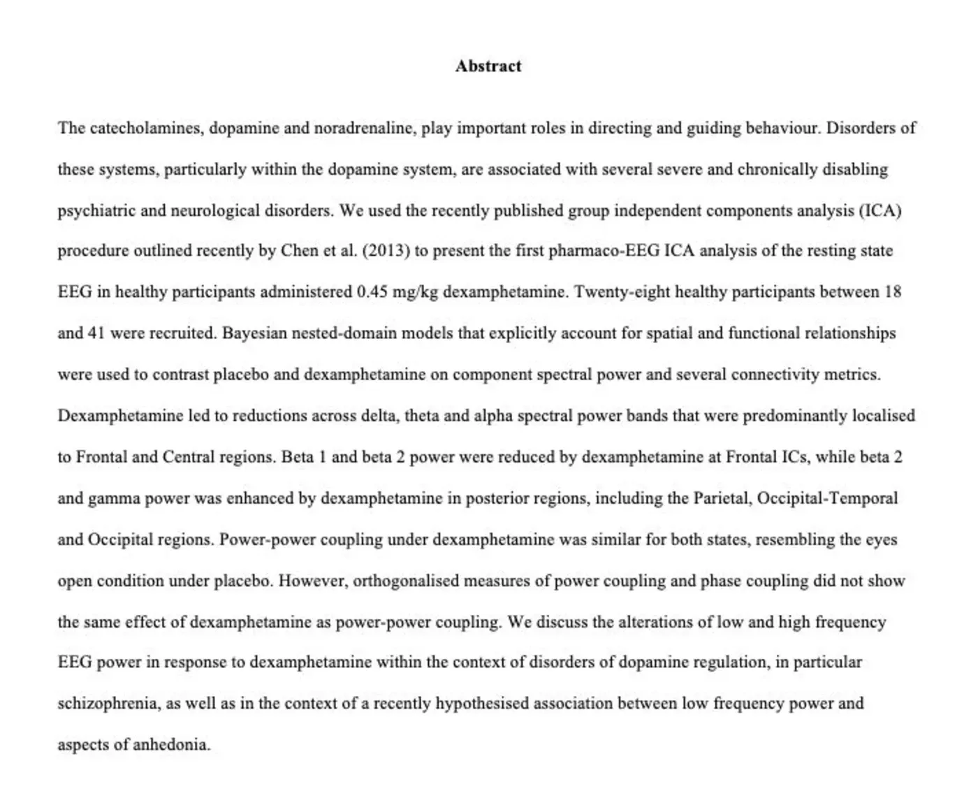
Dopamine and noradrenaline steer behaviour, and disorders of those systems sit under several psychiatric and neurological conditions. This is a pharmaco-EEG study of rest, not of task performance. Twenty-eight healthy adults between 18 and 41 were given 0.45 mg/kg dexamphetamine or placebo.
The approach
Group independent components analysis separates the resting EEG. Bayesian nested-domain models then compare component power and several connectivity metrics, taking the spatial and functional relationships among components into account.
Dexamphetamine reduced delta, theta, and alpha power, mostly in frontal and central components. Frontal beta fell. Posterior beta-2 and gamma rose, including parietal, occipital-temporal, and occipital regions. Power-power coupling under the drug resembled the eyes-open state under placebo. Orthogonalised power coupling and phase coupling did not show that same effect. The discussion places the low- and high-frequency changes next to disorders of dopamine regulation, including schizophrenia, and next to a proposed link between low-frequency power and anhedonia.
- 28 healthy adults, 0.45 mg/kg dexamphetamine versus placebo
- Group ICA of the resting EEG
- Delta, theta, and alpha power down, mostly frontal and central
- Posterior beta-2 and gamma up
- Power-power coupling shifts; phase coupling does not follow it
- Albrecht, Roberts, Price, Lee, Iyyalol, and Martin-Iverson. Human Brain Mapping, 37(2), 570-588
The paper: research-repository.uwa.edu.au .